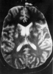
 Figure 1. T2-weighted MRI obtained during the first hospital admission of the patient. Multiple foci of abnormal high signal intensity can be seen within the basal ganglia. The T1-weighted MRI was essentially normal.
|
|
Figure 1. T2-weighted MRI obtained during the first hospital admission of the patient. Multiple foci of abnormal high signal intensity can be seen within the basal ganglia. The T1-weighted MRI was essentially normal.
|